- Visibility 13 Views
- Downloads 3 Downloads
- DOI 10.18231/j.ijpns.2022.006
-
CrossMark
- Citation
Pentalogy of cantrell, type 2- A rare entity
- Author Details:
-
Deepali Bangalia
-
Satyendr Sonkariya *
-
Chandradeep Mastan
-
Dinesh Kumar Barolia
Introduction
Pentalogy of Cantrell or Cantrell syndrome, a rare congenital malformation was first described by Cantrell in 1958.[1] It consists of five midline congenital anomalies, namely of heart, pericardium, diaphragm, sternum and abdominal wall. It can be divided into two types: 1) Complete and 2) Partial. Complete type refers to presence of all five defects while partial Pentalogy of Cantrell doesn’t contain all five defects.[2] The collective defects result from failure of either differentiation or migration of mesenchymal or mesodermal structures during embryonic phase of development.[3]
Case Report
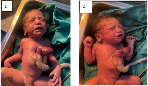
A newborn female child weighing 2.4 kg at birth weight and 46 cm height was admitted at our institute. She had APGAR score 7 at 1 minute and 8 at 5 minutes with normal tone, and cry. There was peripheral cyanosis on clinical examination at the time of admission at NICU Rajkiya Mahila Chikitsalaya just after birth with complain of major congenital malformation of midline supraumbilical, thoracoabdominal wall defect (supra umbilical omphalocele) and evisceration of intestine with complete thoracic ectopic cordis.
There was visible Ectopic heart with absence of pericardium. Heart beat was visible outside the thoracic cavity, at the rate of 156 beats per minute. Respiratory Rate was 68 per minute and saturation in all four limbs without oxygen support was 90 percent. Intercostal retractions were there. An abnormal position of heart was detected on antenatal USG at 28 weeks of gestation. She missed folic acid and calcium supplements during pregnancy. There was no family history of congenital and genetic abnormalities and similar family history related to ectopia cordis. There was no history of smoking, alcohol and drug intake. Fetal Echocardiography was done at 30 weeks and 32 weeks gestation which were suggestive of ectopia cordis (heart located outside the thoracic cavity) with large VSD shunting bidirectional.
Newborn was the product of 23 year old primigravida, who was admitted here with 8 months amenorrhea and complaint of pain abdomen. She was in active labour since 10 days. Baby was born by Normal Vaginal Delivery at 36 weeks of gestation in Rajkiya Mahila Chikitsalaya Ajmer and shifted to NICU. Newborn’s heart was covered with sterile gauze soaked in warm saline. Newborn received intensive medical management. We planned for surgical management but unfortunately child expired on day 4.

Discussion
It is documented that Ectopia cordis was first time observed 5000 years ago.[4] The term ectopia cordis was first time explained by Haller in 1706.[5] The incidence of ectopia cordis is 8 per million live births. It commonly associated with other congenital anomalies like Atrial septal defect, Ventricular septal defect, Tetralogy of Fallot, Tricuspid atresia, Double outlet right ventricle, Non-cardiac malformations, Pentalogy of Cantrell, Omphalocele, Anterior diaphragmatic hernia, Cleft palate.[6], [7], [8] In 1958 Cantrell proposed the syndrome of defects involved omphalocele, sternal defect, diaphragmatic defect, pericardial defect. These all five components named Pentalogy of Cantrell’s.[1] Cantrell’s syndrome was classified in three groups by Toyama WM. Class 1 — all five components of Cantrell’s Pentalogy should present. Class 2 — four components of Cantrell’s Pentalogy may present but intracardiac lesion and ventral abdominal wall abnormalities must present. Class 3 — this class formed by incomplete components of Cantrell’s Pentalogy but sternal defect must present.[9] Our case was presented with omphalocele, ectopia cordis with ventricular septal defect and absent pericardium. According to Toyama classification our case classified in class 2.
Etiology of Cantrell’s Pentalogy is not yet clear. But literature suggests that most of the cases show sporadic pattern and some cases indicate genetic association. Its association with trisomy 18 and 21 and with turners syndrome was reported in literature.[10], [11], [12]
The ventral body wall begins to develop by eight weeks of embryonic life through differentiation and proliferation of mesoderm followed by its lateral migration. The heart originally develops in a cephalic location and reaches its definitive position by the lateral folding and ventral flexing of the embryo at about the 16th–17th day. Midline fusion and Thoracic, abdominal cavity formation is completed by the 9th week of embryonic life. Failure of fusion in mid line mesoderm causes development of Cantrell’s Pentalogy spectrum.[13] Partial and complete Ectopia Cardia with evisceration is dependent on this stage of fusion.
Pentalogy of Cantrell or Ectopic cardiac is diagnosed by routine antenatal USG as early as 10 to 12 weeks of gestation.[14] In our case, ectopic cordis was diagnosed by USG at 26 weeks of gestation. Management of Cantrell’s Pentalogy is the challenge and very difficult task. It is multidisciplinary management. Cutler and Wilens did first attempt to repair the ectopia cordis in 1925.[15] The first successful repair was done in 1975 by Knoop of thoracic ectopic cordis.[16]
Conclusion
Pentalogy of Cantrell is a rare congenital entity, which requires staged surgical management for complete repair. The prognosis of this disease is very poor. However, with advancement in medical science number of survivals after surgical management is increasing. The rarity and unknown etiology is reason to report.
Statement of Ethics
The author confirms that caregivers of their patients were fully informed, and they agreed to report this case.
Source of Funding
None.
Conflict of Interest
The author declares that there is no conflict of interest.
References
- J R Cantrell, J A Haller, M M Ravitch. A syndrome of congenital defects involving the abdominal wall, sternum, diaphragm, pericardium, and heart. Surg Gynecol Obstet 1958. [Google Scholar]
- Mihaela Grigore, Romeo Micu, Roxana Matasariu, Odetta Duma, Anca Lucia Chicea, Radu Chicea. Cantrell syndrome in the first trimester of pregnancy: imagistic findings and literature review. Med Ultrason 2020. [Google Scholar] [Crossref]
- Adele P Williams, Raoud Marayati, Elizabeth A Beierle. Pentalogy of Cantrell. Semin Pediatr Surg 2019. [Google Scholar] [Crossref]
- H B Taussig. World survey of the common cardiac malformations: developmental error or genetic variant?. Am J Cardiol 1982. [Google Scholar] [Crossref]
- T Geva, S Van-Praagh, R Van-Praagh. Thoracoabdominal ectopia cordis with isolated infundibular atresia”. Am J Cardiol 1990. [Google Scholar] [Crossref]
- Myung K Park. Pediatric Cardiology for Practitioners. 2008. [Google Scholar]
- J J Amato, W I Douglas, U Desai, S Burke. Ectopia cordis. Chest Surg Clin N Am 2000. [Google Scholar]
- Daniel Bernstein. Kliegman: Nelson Textbook of Pediatrics. 2011. [Google Scholar]
- W M Toyama. Combined congenital defects of the anterior abdominal wall, sternum, diaphragm, pericardium, and heart: A case report and review of the syndrome. Pediatrics 1972. [Google Scholar]
- C R King. Ectopia cordis and chromosome errors. Pediatrics 1980. [Google Scholar]
- S P Soper, L R Roe, H E Hoyme, J J Clemmons. Trisomy 18 with ectopia cordis, omphalocele, and ventricular septal defect: case report. Pediatr Pathol 1986. [Google Scholar] [Crossref]
- Feico J J Halbertsma, Anton van Oort, Frans van der Staak. Cardiac diverticulum and omphalocele: Cantrell's pentalogy or syndrome. Cardiol Young 2002. [Google Scholar] [Crossref]
- Scott A Engum. Embryology, sternal clefts, ectopia cordis, and Cantrell’s pentalogy. Semin Pediatr Surg 2008. [Google Scholar] [Crossref]
- Cuillier F, Avignon MS, A Avignon. Pentalogy of Cantrell, 11 weeks. Pentalogy of Cantrell, 11 weeks © Cuillier 2006. [Google Scholar]
- George David Cutler, Gustav Wilens. Ectopia cordis: a report of a case. Am J Dis Child 1925. [Google Scholar] [Crossref]
- N C Saxena. Ectopia cordis child surviving: prostheis fails. Pediatr News 1976. [Google Scholar]
